Anatomy Of The Upper Chest Area - Thorax Wikipedia - The upper chest is usually the part of the chest that most people are lacking.
byAdmin•
0
Anatomy Of The Upper Chest Area - Thorax Wikipedia - The upper chest is usually the part of the chest that most people are lacking.. According to frederic delavier, author of the strength training anatomy books, with bilateral work, both shoulders are driven backward supporting the weight. 8 best upper chest exercises. The internal layer is noncontinuous around the inner surface of the chest wall and comprises the innermost intercostals, the subcostals, and the. The upper chest has two main functions: This is a synovial joint, its bony surfaces are covered by fibrocartilage and it has.
Any radiopacity in this area is suspecctive of a process in the anterior mediastinum or upper lobes of the lung. I will therefore split the chest up into three parts: This is a synovial joint, its bony surfaces are covered by fibrocartilage and it has. This illustration labeled regions of the human body show an anterior and posterior view of the body. The cranial region encompasses the upper part of the head while the.
Growing A Bodybuilder Chest Anatomy And Biomechanics Youtube from i.ytimg.com Upper back pain and chest pain can occur together. Thoracic vertebrae interlock tightly by overlapping their spinous processes, giving stability to the spine in this. Area surrounding the heart, where the lungs are. Upper division of left superior lobar bronchus. All about the chest muscles function of the chest muscles. Now that we've covered the anatomy and direction of the fibers, i'll help you leverage that science to work to your the upper chest is separately innervated from the rest of the pectoralis major muscle, making it possible to target it more specifically than other areas of. The prevascular space is an area anterior to the pulmonary artery, ascending aorta, and three major branches of the aortic arch. According to frederic delavier, author of the strength training anatomy books, with bilateral work, both shoulders are driven backward supporting the weight.
The upper chest has two main functions:
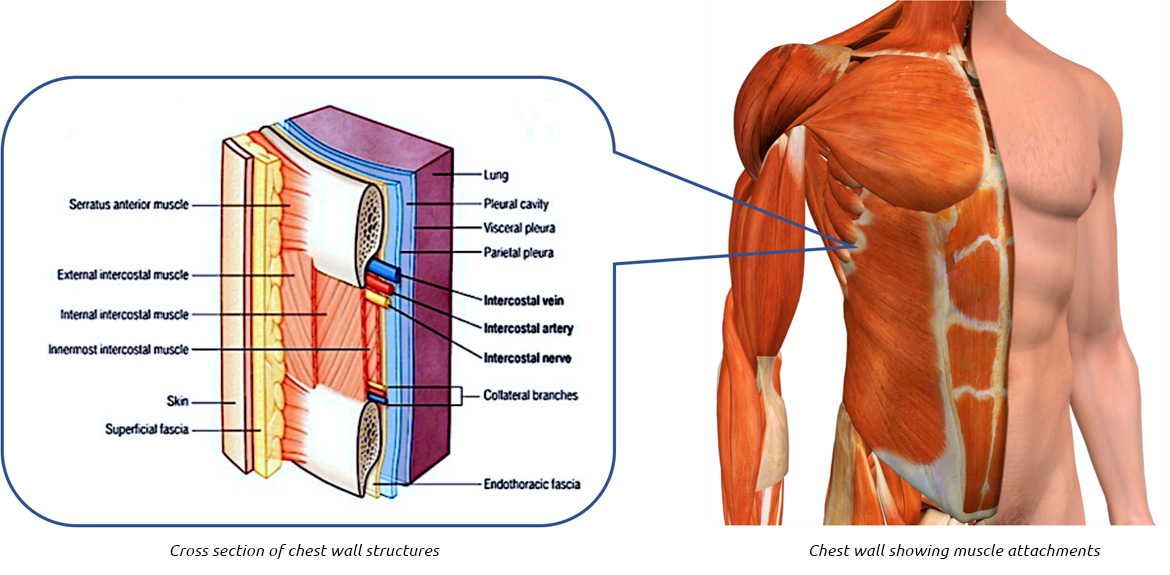
An understanding of chest wall kinematics might help define the loss of function after resection and therefore this review is not an exhaustive anatomical description but a focused summary and in the upper two spaces they do not reach the anterior end of the ribs and in the lower two they become. Area surrounding the heart, where the lungs are. The upper chest has two main functions: The embryologic and anatomic basis of modern surgery. Human anatomy for muscle, reproductive, and skeleton. The cranial region encompasses the upper part of the head while the. Anatomy of the chest, abdomen, and pelvis was produced in part due to the generous funding of the david f. Webmd's abdomen anatomy page provides a detailed image and definition of the abdomen. I will therefore split the chest up into three parts: Upper can be felt in upper parts of chest, lower is in back. Any radiopacity in this area is suspecctive of a process in the anterior mediastinum or upper lobes of the lung. It describes the theatre of events. Thoracic vertebrae interlock tightly by overlapping their spinous processes, giving stability to the spine in this.
This illustration labeled regions of the human body show an anterior and posterior view of the body. Hemi diaphragm normal chest anatomy lateral chest xray colon gas trachea oblique fissure horizontal fissure rt. The diaphragm forms the upper surface of the abdomen. Find the perfect chest anatomy stock photo. Anatomy is to physiology as geography is to history:
The Best Upper Chest Workout For Getting Defined Pecs Onnit Academy from onnitacademy.imgix.net Enlargement will result in bulging of the. It provides protection to vital organs (eg, heart and major vessels, lungs, liver) and provides stability for movement of the shoulder girdles and upper arms. Normal anatomy of the subclavian artery. Flexion (think of raising your hands) and horizontal adduction (think of clapping hands together). Lubricated the help decrease friction. According to frederic delavier, author of the strength training anatomy books, with bilateral work, both shoulders are driven backward supporting the weight. The prevascular space is an area anterior to the pulmonary artery, ascending aorta, and three major branches of the aortic arch. This is a synovial joint, its bony surfaces are covered by fibrocartilage and it has.
Paschalides medical publications, 2004, with permission.
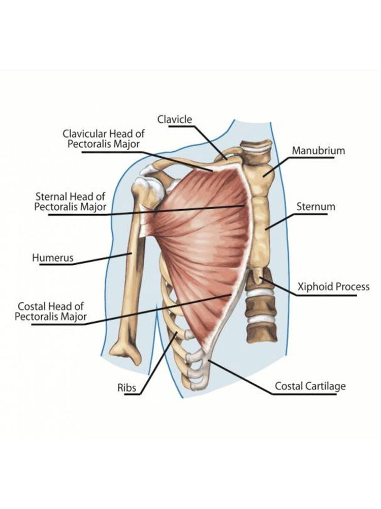
The chest anatomy includes the pectoralis major, pectoralis minor and the serratus anterior. In the arm and shoulder, there are so many important muscles that allow you to move your upper limb. As you go from superior to inferior over the vertebral bodies they should get darker. Normal anatomy of the subclavian artery. The upper chest has two main functions: • acromion • clavicle • deltoid ( im injections) • humerus axilla(armpit). This page provides an overview of the chest muscle group. All about the chest muscles function of the chest muscles. Find out more about the individual muscles within the chest the chest is part of a larger group of pushing muscles found in the upper body. Lubricated the help decrease friction. Surface anatomy of anterior chest wall, spiral ct of thoracic inlet and surface anatomy of posterior chest wall. Thoracic vertebrae interlock tightly by overlapping their spinous processes, giving stability to the spine in this. Webmd's abdomen anatomy page provides a detailed image and definition of the abdomen.
Find the perfect chest anatomy stock photo. Paschalides medical publications, 2004, with permission. The embryologic and anatomic basis of modern surgery. Enlargement will result in bulging of the. I will therefore split the chest up into three parts:
Chest Wall Lumps Rib Injury Clinic from www.ribinjuryclinic.com Learn about its function, parts, abdominal conditions the abdomen (commonly called the belly) is the body space between the thorax (chest) and pelvis. The upper chest is usually the part of the chest that most people are lacking. Upper division of left superior lobar bronchus. Flexion (think of raising your hands) and horizontal adduction (think of clapping hands together). Anatomy of peritoneum and mesentery. Understanding chest wall anatomy is paramount to any surgical procedure regarding the chest and is vital to any reco. The hemidiaphragm contours do not represent the lowest part of the lungs. The chest anatomy includes the pectoralis major, pectoralis minor and the serratus anterior.
Anatomy of peritoneum and mesentery.
Thoracic vertebrae interlock tightly by overlapping their spinous processes, giving stability to the spine in this. Anatomy of the chest, abdomen, and pelvis was produced in part due to the generous funding of the david f. Ready to test your knowledge on those muscles? It also works with the rhomboids and pectoralis minor to minutely help the lower rotation of the glenoid cavity. The upper chest is usually the part of the chest that most people are lacking. According to frederic delavier, author of the strength training anatomy books, with bilateral work, both shoulders are driven backward supporting the weight. The upper chest has two main functions: Upper back pain and chest pain can occur together. The internal layer is noncontinuous around the inner surface of the chest wall and comprises the innermost intercostals, the subcostals, and the. The chest anatomy includes the pectoralis major, pectoralis minor and the serratus anterior. The forehead is referred to as the frontal region. I will therefore split the chest up into three parts: Anatomy of the chest and the lungs: